Back Human Bones Labeled - Human Skeleton Front Back Main Parts Stock Vector Royalty Free 1092554153 - Your skeleton can be divided into two main parts.
Back Human Bones Labeled - Human Skeleton Front Back Main Parts Stock Vector Royalty Free 1092554153 - Your skeleton can be divided into two main parts.. The occiput (co), also known as the occipital bone, is a flat bone that forms the back of the head. The remaining small bones or ossicles below the sacrum are also fused together and called the tailbone or coccyx. Human musculature bodybuilding infographic muscular system vector human anatomy back muscle anatomy bicep male muscular anatomy human body anatomy female female anatomy muscle hamstrings muscle. The muscles of the lower back help stabilize, rotate, flex, and extend the spinal column, which is a bony tower of 24 vertebrae that gives the body structure and houses the spinal cord. The vertebral column of the lower back includes the five lumbar vertebrae, the sacrum, and the coccyx.
Super angebote für rag n bone human hier im preisvergleich. Using this atlas of human anatomy of the spine and back. When humans are born we have close to 300 bones, and over time they fuse together. Huge collection, amazing choice, 100+ million high quality, affordable rf and rm images. All the other bones in the skull are firmly attached to one another by.
File Human Skeleton Back En Svg Wikipedia from upload.wikimedia.org The remaining small bones or ossicles below the sacrum are also fused together and called the tailbone or coccyx. It runs down the centre of the body. The number of bones in the human body at birth is 300. When most people mention their back, what they are actually referring to is their spine. Related posts of back bone anatomy body anatomy bones. This article is concerned primarily with the gross structure and the function of the skeleton of the normal. The axial skeleton is made up of the skull, backbone, breastbone, and ribs. The vertebral column is the defining characteristic of a vertebrate in which the notochord (a flexible rod of uniform composition) found in all chordates has been replaced by a segmented series of bone:
The bottom of the spine is called the sacrum.
It is the surface of the body opposite from the chest and the abdomen. Super angebote für rag n bone human hier im preisvergleich. The most common variations include sutural (wormian) bones, which are located along the sutural lines on the back of the skull, and sesamoid bones which develop within some tendons, mainly in the hands and feet. The human back, also called the dorsum, is the large posterior area of the human body, rising from the top of the buttocks to the back of the neck. Everything else that hangs from this, like the arms, legs, shoulders, and hips, is called the appendicular skeleton. It is made up of several vertebral bodies usually fused together as one. That is the joint connecting the lower jaw, or mandible, to the rest of the skull. The back functions are many, such as to house and protect the spinal cord, hold the body and head upright, and adjust the movements of the upper and lower limbs. All the other bones in the skull are firmly attached to one another by. This article is concerned primarily with the gross structure and the function of the skeleton of the normal. Related posts of back bone anatomy body anatomy bones. When humans are born we have close to 300 bones, and over time they fuse together. The vertebral column of the lower back includes the five lumbar vertebrae, the sacrum, and the coccyx.
It is the surface of the body opposite from the chest and the abdomen. Overview of bones & the axial skeleton. The most common variations include sutural (wormian) bones, which are located along the sutural lines on the back of the skull, and sesamoid bones which develop within some tendons, mainly in the hands and feet. It is designed to be incredibly strong, protecting the highly sensitive nerve roots, yet highly flexible, providing for mobility on many different planes. They also provide for the attachment of muscles, and help us move around.
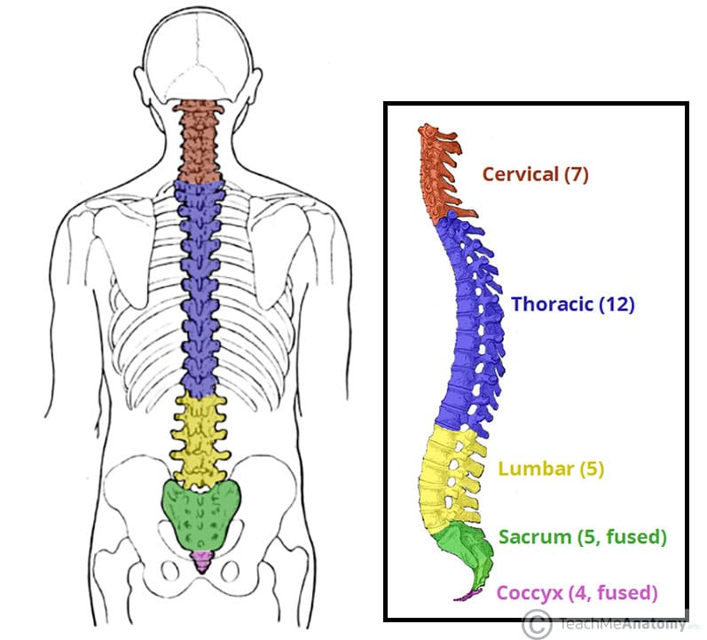
The Vertebral Column Joints Vertebrae Vertebral Structure from teachmeanatomy.info Bones of the pelvis and lower back the bones of the pelvis and lower back work together to support the body's weight, anchor the abdominal and hip muscles, and protect the delicate vital organs of the vertebral and abdominopelvic cavities. Super angebote für rag n bone human hier im preisvergleich. The occiput (co), also known as the occipital bone, is a flat bone that forms the back of the head. Nerves in your lower back five pairs of lumbar spinal nerves labeled l1 to l5 branch off your spinal cord and exit through small holes between the vertebrae. The human skeleton anatomy chart shows three views of the human skeleton (front, back and side) and is painstakingly labeled and painted, producing one of the most captivating and beautiful anatomical charts available. Human body anatomy female female anatomy muscle shoulder blade pain anatomy back muscles bones man female anatomy body muscles in a body female anatomy muscole shoulder concept muscular sysyem. Spinal anatomy is a remarkable combination of strong bones, flexible ligaments and tendons, large muscles and highly sensitive nerves. That is the joint connecting the lower jaw, or mandible, to the rest of the skull.
No need to register, buy now!
This anatomy chart is ideal for higher education or patient consultation. There is only one movable joint in the skull. This includes the head, facial, hyoid, auditory, trunk, ribs, and sternum. Body anatomy bones 12 photos of the body anatomy bones anatomy all bones body, anatomy bones human body. The spine runs from the base of your skull down the length of your back, going all the way down to your pelvis. Some individuals may also have additional (i.e., supernumerary) cervical ribs or lumbar vertebrae. That is the joint connecting the lower jaw, or mandible, to the rest of the skull. Human body anatomy female female anatomy muscle shoulder blade pain anatomy back muscles bones man female anatomy body muscles in a body female anatomy muscole shoulder concept muscular sysyem. The back functions are many, such as to house and protect the spinal cord, hold the body and head upright, and adjust the movements of the upper and lower limbs. The bones of the superior portion of the skull are known as the cranium and protect the brain from damage. Human back bone chart, find out more about human back bone chart. The human skeleton anatomy chart shows three views of the human skeleton (front, back and side) and is painstakingly labeled and painted, producing one of the most captivating and beautiful anatomical charts available. On anatomical parts the user can choose to display the various structures in colored illustrations of the anatomy of the back and spine:
See human back anatomy stock video clips. Overview of bones & the axial skeleton. Find the perfect anatomy rear view back human stock photo. Nerves in your lower back five pairs of lumbar spinal nerves labeled l1 to l5 branch off your spinal cord and exit through small holes between the vertebrae. Human skeleton, the internal skeleton that serves as a framework for the body.
Skeletal System from claypoole.ucfsd.org Find the perfect anatomy rear view back human stock photo. Each lumbar spinal level is numbered from top to bottom—l1 through l5, or l6. The muscles of the lower back help stabilize, rotate, flex, and extend the spinal column, which is a bony tower of 24 vertebrae that gives the body structure and houses the spinal cord. The bones of the skull. There is only one movable joint in the skull. The number of bones in the human body at birth is 300. It is designed to be incredibly strong, protecting the highly sensitive nerve roots, yet highly flexible, providing for mobility on many different planes. The human skeleton anatomy chart shows three views of the human skeleton (front, back and side) and is painstakingly labeled and painted, producing one of the most captivating and beautiful anatomical charts available.
It also covers some common conditions and injuries that can affect the back.
This framework consists of many individual bones and cartilages.there also are bands of fibrous connective tissue—the ligaments and the tendons—in intimate relationship with the parts of the skeleton. On anatomical parts the user can choose to display the various structures in colored illustrations of the anatomy of the back and spine: The back functions are many, such as to house and protect the spinal cord, hold the body and head upright, and adjust the movements of the upper and lower limbs. The bones provide a structural framework and protection to the soft organs. The part of the nerve that emerges out of the spine is called the nerve root. The spine runs from the base of your skull down the length of your back, going all the way down to your pelvis. Related posts of back bone anatomy body anatomy bones. Human body anatomy female female anatomy muscle shoulder blade pain anatomy back muscles bones man female anatomy body muscles in a body female anatomy muscole shoulder concept muscular sysyem. This vertebra supports the skull. This anatomy chart is ideal for higher education or patient consultation. Primarily, they are referred to as long or short. Human musculature bodybuilding infographic muscular system vector human anatomy back muscle anatomy bicep male muscular anatomy human body anatomy female female anatomy muscle hamstrings muscle. Overview of bones & the axial skeleton.
The part of the nerve that emerges out of the spine is called the nerve root human back bones. That is the joint connecting the lower jaw, or mandible, to the rest of the skull.


0 Komentar